
Welcome to the new Gustave Roussy website
A new website designed to offer you a simpler, clearer, and more intuitive experience. Patients, caregivers, healthcare professionals, and donors: find information, news, and services more easily.
Skin Cancer
Directors:
Prof. Caroline Robert, Head of the Dermatology Department;
Dr. Émilie Routier, Chair of the Dermatological Pathology Committee
Contact
Tel.: +33 (0)1 42 11 46 88

Understanding the Disease
Skin cancer is an abnormal and uncontrolled proliferation of skin cells, most often triggered by excessive exposure to ultraviolet (UV) radiation from the sun or tanning beds.
There isn’t just one type of skin cancer—there are several.
Carcinomas, which are the most common types of skin cancer, generally have a good prognosis.
Squamous cell carcinoma: It develops from the keratinocytes of the epidermis. It can occur spontaneously, but is often preceded by a precancerous skin or mucosal lesion that progresses in several stages (actinic keratosis, squamous cell carcinoma in situ, also known as Bowen’s disease). It can present in various forms and affect the oral or genital mucous membranes.
Squamous cell carcinoma has a good prognosis, but its management must be rigorous because it can recur locally or lead to metastases. Metastases occur when cancer cells have spread along lymphatic pathways (to the lymph nodes) or through the bloodstream to other organs. For forms with a poor prognosis and recurrences, treatment requires discussion and a decision made during a multidisciplinary team meeting (MDT).
Basal cell carcinoma (BCC): It develops from keratinocytes in the deep layer of the epidermis. It occurs without a preexisting lesion. There are several clinical forms: nodular BCC, superficial BCC, and sclerodermoid BCC. BCC does not metastasize, but it must be treated rigorously because it can cause significant local damage. Its progression is slow, and treatment can be approached without haste.
Merkel cell neuroendocrine carcinoma (CCM): This is a rare and aggressive cutaneous carcinoma with a high risk of recurrence, and metastatic spread can occur anywhere. Most relapses occur within two years of the primary tumor’s discovery. In 2008, scientists discovered the Merkel cell polyomavirus, a human virus present in approximately 80% of MCC cases. Most people have already been exposed to polyomaviruses (members of a family of double-stranded DNA viruses) by the age of 20. When the Merkel cell polyomavirus infects a cell, it produces proteins that can lead to uncontrolled cell growth, resulting in cancer.
These cancers are rarer but more aggressive because they can spread rapidly throughout the body. They develop from melanocytes. These melanocytes produce melanin and are responsible for our skin color. Melanoma most commonly affects patients around age 50, but it can be diagnosed in younger individuals. However, it is extremely rare before puberty. It primarily appears on the skin, but it can also affect the mucous membranes or the eye (choroid or conjunctiva).
Histological analysis confirms the diagnosis, determines the thickness of the melanoma (Breslow index), and assesses the degree of skin invasion, thereby helping to determine the appropriate follow-up treatment. Its thickness is directly correlated with the prognosis: the thicker the melanoma, the greater the risk of metastasis.
Melanoma by the Numbers
-
17,922
new cases in 2023
-
62 years old
average age at diagnosis among women
-
68 years old
average age at diagnosis among men
Risk Factors
Acute exposure: Repeated sunburns, especially during childhood, are a major risk factor.
Chronic exposure: Prolonged and cumulative exposure to the sun throughout one’s lifetime also increases the risk.
Artificial UV radiation: Using tanning beds is associated with an increased risk, particularly before age 35.
Light phototype (I and II): People with fair skin, red or blond hair, and light-colored eyes are at significantly higher risk.
Atypical nevus syndrome: The presence of a large number of irregular-looking "moles" is a factor requiring close monitoring.
Congenital nevi: Present at birth, their risk of becoming malignant increases with size.
Family history: About 10% of melanomas occur in a family context (at least two cases in the family), particularly in connection with mutations in the CDKN2A/p16, CDK4, or MITF genes.
Xeroderma pigmentosum: a rare genetic disorder that impairs the skin’s ability to repair UV-induced damage, resulting in a very high risk.
Immunosuppression: A weakened immune system (organ transplant, HIV, immunosuppressive treatments) reduces the body’s ability to eliminate abnormal cells.
Screening
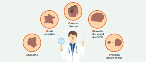
Melanoma screening relies primarily on observing the skin and detecting suspicious lesions early.
To identify suspicious melanomas or moles (naevi), the ABCDE acronym is used:
- A: asymmetry of the lesion
- B: irregular, notched borders
- C: uneven color ranging from brown to black, possibly with depigmented, white, or flesh-colored areas
- D: diameter greater than 6 mm (non-specific)
- E: This is the key criterion, characterized by rapid progression—usually within less than 6 months—and changes in size, shape, texture, and color.
In patients with a large number of pigmented nevi (moles), the suspicious lesion differs from the others, which generally resemble one another (the “ugly duckling” sign).
Coverage
The Dermatology Committee at Gustave Roussy treats patients with skin cancer and those experiencing skin-related side effects from cancer treatments.
This care begins with an initial evaluation.
Initial Evaluation of Skin Cancers
The evaluation is based primarily on a clinical examination and a search for other suspicious skin lesions and/or signs of distant metastasis. For carcinomas, imaging studies such as CT or MRI scans are performed only in cases of large tumors, recurrent tumors, or tumors located in high-risk areas such as the midface.
For cancers that may metastasize, imaging studies are not mandatory; however, an initial evaluation is recommended to facilitate monitoring by assessing the current status and documenting any potential abnormalities, such as hepatic angiomas, renal cysts, or sequelae of tuberculosis…
In cases of lymph node involvement, an evaluation to detect distant metastases via chest X-ray and abdominal ultrasound or CT scan is recommended.
The InstaDiag Skin Cancer Assessment Pathway
To reduce wait times for care, optimize the care pathway, and advance research, Gustave Roussy operates a skin cancer center. This program is divided into two pathways: the first is an outpatient program for the most superficial lesions, and the second, called Instadiag Skin Cancer, is dedicated to more complex lesions.
→ Learn more about the Instadiag pathway [note: link to be inserted to the Instadiag section]

Treatments
The treatments offered to patients with skin cancer depend on the type and stage of their tumor.
Surgery
The primary treatment for melanoma is surgery. First, the surgeon removes the tumor along with a safety margin around it. The lesion is then analyzed in a laboratory, in particular to measure its thickness—a key piece of information for determining the next steps in treatment. A second procedure is then performed to ensure that all edges of the excised area are clear of cancer.
In the case of distant metastases
If there is a single, operable metastasis, surgery may be considered. In this case, it will be followed by additional (adjuvant) treatment with immunotherapy.
In cases of inoperable metastases or multiple metastases, drug therapy must be initiated. First, testing for a genetic mutation in the BRAF gene must be performed.
If a BRAF mutation is detected, targeted therapy combining two drugs (anti-BRAF and anti-MEK) may be recommended. Immunotherapy may also be recommended. Radiation therapy is primarily recommended for brain and bone metastases. Stereotactic radiation therapy yields good results for certain brain metastases.
The first-line treatment is surgery. The squamous cell carcinoma must be completely removed, along with margins of healthy tissue surrounding the tumor. Reconstruction may therefore be necessary afterward.
Surgery is always the treatment of choice for invasive basal cell carcinomas.
When surgery is not an option, a targeted therapy called vismodegib (an oral medication) has shown good results. Radiation therapy may also be recommended.
Surgical treatment of the primary tumor is always the preferred option, followed by radiation therapy to the surgical site. In advanced or metastatic cases, immunotherapy with an anti-PD-L1 agent (Avelumab) may be recommended. An alternative to this treatment may be chemotherapy with carboplatin and etoposide.
In cases where standard treatment has proven ineffective, and if the patient wishes, they can often be offered the opportunity to participate in a clinical trial if they are eligible. This allows them to receive innovative treatments that are not yet available on the market.
Search
The Dermatology Committee, through Professor Caroline Robert, is particularly involved in research aimed at identifying the mechanisms of resistance to melanoma treatments, to the identification of biomarkers of efficacy and resistance to therapies, and to the development of new therapeutic strategies.
This research is being conducted within the “Predictive Biomarkers and New Molecular Strategies in Cancer Therapy” team. The research is structured around three main areas:
- Identification of mechanisms of resistance to melanoma treatments
- Identification of biomarkers of efficacy and resistance
- Development of new therapeutic strategies
- New Antitumor Therapies
For several years now, clinical trials have demonstrated the efficacy of new therapies for advanced, inoperable, or metastatic skin cancers. Two major approaches have received marketing authorization (MA) in France:
- Targeted therapies
- BRAF inhibitors: targeting a genetic mutation found in about half of all melanoma cases.
- Immunotherapies
- Anti-CTLA-4: ipilimumab
- Anti-PD-1: nivolumab, pembrolizumab
- Anti-PDL-1: avelumab
Ongoing clinical trials
Several clinical trials are currently underway for these innovative treatments, including:
- Combinations of targeted therapies and immunotherapy
- New drugs or drug combinations
Join a clinical trial
Many clinical trials are open to patients with cancer. Participating in a clinical trial provides access to innovative treatments while contributing to the development of new therapies.

Contact
Head of the Dermatology Department, Prof. Caroline Robert
Chair of the Dermatological Pathology Committee: Dr. Émilie Routier
Contact: +33 (0)1 42 11 46 88
Appointments and Second Opinions

Testimonials
Odile’s Story: Her Experience Being Treated for Skin Cancer at Gustave Roussy
“I received a wonderful welcome at Gustave Roussy when I was diagnosed with the disease. I received truly personalized care that restored my self-confidence and helped me believe I could recover.”

-
Cancer et soleil : les bons réflexes à adopter
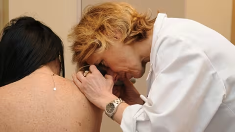
L’été est trop souvent synonyme d’exposition excessive au soleil, dont les rayonnements ultraviolets sur la peau sont responsables chaque année en France de plus de 10 000 nouveaux cas de mélanomes, selon les données de Santé Publique France. Se protéger soi-même et ses proches de façon optimale est un enjeu majeur de santé publique, d’autant plus pour les patients traités ou guéris d’un cancer, chez qui les rayonnements UV peuvent engendrer de nombreuses complications.
-
Dix ans d’avancées scientifiques dans les traitements du mélanome
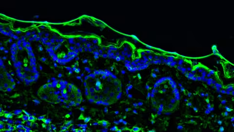
Le traitement des mélanomes métastatiques a considérablement progressé ces dernières années grâce aux immunothérapies et traitements ciblés.


Frequently Asked Questions
Skin cancer is an abnormal proliferation of skin cells. There are several types, the most common being carcinomas (basal cell and squamous cell) and melanoma, which is the most serious because it can spread rapidly to other organs.
Exposure to ultraviolet radiation (sunlight, tanning beds) is the main cause. Other factors also play a role: fair skin, family history, a large number of moles, or a weakened immune system.
We use the ABCDE rule: a lesion that is asymmetrical, has irregular borders, is uneven in color, is larger than 6 mm in diameter, and—most importantly—is changing rapidly should raise concern and prompt a medical consultation.
By limiting sun exposure during the hottest hours of the day, wearing protective clothing, applying high-SPF sunscreen, and avoiding tanning beds.
When detected early, skin cancer—including melanoma—can be successfully treated in the vast majority of cases. That is why early detection and regular monitoring of your skin are essential.