
Welcome to the new Gustave Roussy website
A new website designed to offer you a simpler, clearer, and more intuitive experience. Patients, caregivers, healthcare professionals, and donors: find information, news, and services more easily.
Department of Medical Imaging
Department Chair
Prof. Corinne Balleyguier
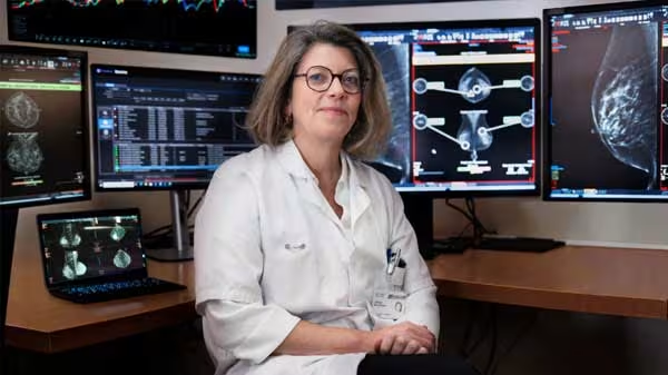
Medical imaging plays a vital role at every stage of cancer care: diagnosis, assessment of the extent of the disease, preparation for treatment, and monitoring of treatment effectiveness.
At Gustave Roussy, the diagnostic imaging, nuclear medicine, and endocrine oncology teams rely on state-of-the-art equipment to provide patients with accurate tests tailored to their individual situations.
Take it from an expert
Prof. Corinne Balleyguier
Head of the Department of Medical Imaging
“Imaging has become an essential tool in precision medicine. Our goal is to provide every patient with the most reliable information to guide treatment decisions and evaluate the effectiveness of treatments throughout their care journey.”

Technical Facilities
-
2
scanners, including 1 photonic scanner
-
2
MRI (+ 1 currently being installed)
-
2
PET Scan
-
2
gamma cameras
-
3
mammography machines
-
1
dedicated radiology room
Our Areas of Expertise
Diagnostic Imaging Department
The department performs imaging studies to detect and characterize tumors, assess their extent, and monitor the effectiveness of treatments.
The main imaging modalities used are:
- Ultrasound
- CT scan
- MRI
- Mammography
- Breast tomosynthesis
- Angiomammography
Nuclear Medicine Department
The department uses radioactive substances for diagnostic or therapeutic purposes to obtain functional information about tumors.
The tests performed include, among others:
- Scintigraphy
- PET Scan
- Therapies using radioactive isotopes
Endocrine Oncology
The department provides specialized care for:
- Thyroid cancers
- Adrenal gland tumors
- Neuroendocrine tumors
What is nuclear medicine?
Nuclear medicine is a medical specialty that uses small amounts of radioactive substances to visualize certain bodily functions or treat certain tumors. It often provides additional information to complement that obtained from CT or MRI scans.
Exams Offered
A test that uses X-rays to produce detailed images of the body.
A technique that uses a magnetic field to accurately visualize organs and tissues.
An ultrasound-based examination, frequently used to evaluate certain masses or guide biopsies.
The gold standard imaging test for evaluating breast abnormalities.
A nuclear medicine test used to observe metabolic activity in tissues and assess the extent of certain cancers.
A functional test used to assess the functioning of certain organs or tissues.
Research and Innovation
The teams are involved in numerous research projects aimed at improving the accuracy of diagnoses and the evaluation of treatments.
Diagnostic Imaging
- Evaluation of Breast Tomosynthesis
- Development of Spectral Imaging in ENT
- Evaluation of whole-body MRI and functional MRI protocols
- Development of angiomammography
- New Criteria for Evaluating Treatment Response in Clinical Research
Nuclear Medicine
- Introduction of new isotopes
- Participation in innovative therapeutic trials
Endocrine Oncology
- Participation in innovative therapeutic trials
- Contribution to large multicenter cohorts supported by INCa
Management Team
- Prof. Corinne Balleyguier, Department Chair and Head of the Diagnostic Imaging Service
- Dr. Désirée Deandreis, Chief of the Nuclear Medicine Division
- Dr. Éric Baudin, Chief of the Endocrine Oncology Unit
Coordination
- Simon Corbin, Medical-Technical Coordinator
- Marie-Stéphanie Bour, Deputy Management Executive
- Christine Gaillard, Medical Secretariat Manager
Contacts
Department of Medical Imaging
Administrative Office Tel.: +33 (0)1 42 11 43 99
Diagnostic Imaging Department
- Main Office: +33 (0)1 42 11 48 73
- Healthcare Manager: +33 (0)1 42 11 45 09
Department of Nuclear Medicine and Endocrinology
- New Patients: +33 (0)1 42 11 57 94
- Main Office: +33 (0)1 42 11 60 95
- Head Nurse: +33 (0)1 42 11 42 68
Join us
Are you interested in a career in medical imaging?
Imaging teams are regularly hiring!
For any questions regarding opportunities within the Department of Medical Imaging: